noscript

About Electrocardiograms (EKG/ECG)

Learn More About Electrocardiograms (EKG/ECG):

Electrocardiograms (ECG or EKG)
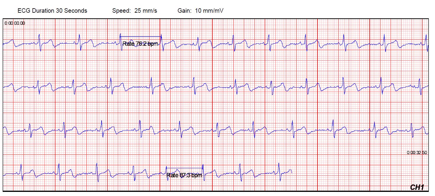
An electrocardiogram (ECG or EKG) is a non-invasive medical test that’s crucial in the field of cardiology for assessing the electrical activity of the heart. This test involves the use of electrodes placed on the skin’s surface to record the heart’s electrical impulses, providing valuable information about the heart’s rhythm, rate, and overall function. ECG is an essential tool for diagnosing and monitoring various heart conditions, and its history, principles, and applications are subjects of great significance in the medical world.
EKG Frequently Asked Questions (FAQs)

Want to learn more about EKGs?

Visit our dedicated FAQ section on the topic.
 

  • EKG
  • GEMS™ Win
Expand All | Collapse All
  • 1. EKG vs ECG: What is the difference?
     

    "EKG" and "ECG" refer to the same medical test: the electrocardiogram. The difference between the two terms lies primarily in the language and terminology used in different regions.

    ECG (Electrocardiogram): This term is more commonly used in European countries and in the international medical community. "ECG" is derived from the German term "Elektrokardiogramm."

    EKG (Electrocardiogram): This term is predominantly used in the United States. The "K" in EKG comes from the German word "Kardiogramm." The use of "K" instead of "C" is attributed to the influence of the German word.

    In essence, an ECG or EKG is a diagnostic test that records the electrical activity of the heart over a period of time. It is commonly used to assess the heart's rhythm and to diagnose various heart conditions.

    Both terms, ECG and EKG, are widely understood in the medical field, and the choice of which term to use often depends on regional conventions and preferences. The test itself involves placing electrodes on the skin to detect and record the electrical impulses produced by the heart as it beats.
     

    Learn more about EKGs
     

    More
  • 2. What is the relationship between a heart monitor and an EKG?
     

    "Heart monitor" is a general term for a device used to monitor the electrical activity of the heart. It can include various types of devices, such as Holter monitors, event monitors, implantable cardiac monitors (ICMs), wearable tech and mobile EKG devices that provide medical-grade heart readings for home use by patients.

    An EKG is a common tool used in heart monitoring. It conducts a specific test that records the electrical activity of the heart over a short period. It produces a visual representation of the heart’s electrical impulses, which is called an electrocardiogram.

    More
  • 3. GEMS™ Components and Setup
     

    The GEMS™ software suite can be set up for many different configurations and networking options. A GEMS™ installation can range from a single PC or be installed in a multi-workstation, networked environment.

    Listed below are the typical components of a GEMS installation and setup.
     

    Component Type Description
    GEMS™ Software (CD) GEMS software can be installed on either single or multiple networked workstations. GEMS is also available in several different versions to meet your specific monitoring needs.
    GEMS™ - Database MS Access or MS SQL The GEMS Database is where all your GEMS patient and physician data is stored. GEMS can either use MS Access or MS SQL formats for its database structure. The Database structure is created through the GEMS program. Databases can be stored locally or on a network, depended on the version purchased.
    GEMS™ - ECG Warehouse File Storage Location The GEMS ECG Warehouse is the set location of where your GEMS ECG files are stored and viewed from. On a networked GEMS environment, the ECGS are usually stored on a server with the GEMS Database.
    ECG Server Service Installed Service (multi-workstation setups only) The ECG Server Service runs as a Service on the workstation or Server that stores the GEMS ECG Warehouse. This service broadcasts the ECG warehouse location to all GEMS PCs throughout your GEMS network. This allows for centralized storage of all ECGs.
    Hardware License Dongle Hardware (USB or Parallel Port) GEMS licensing is controlled by the use of Hardware License Dongles that attaches to each GEMS workstation and are required in order for GEMS to operate. GEMS license dongles are available in either FULL or EDIT versions.
    * EDIT versions cannot receive/record ECG transmissions.
    Telephone Logger Patch (TLP) Hardware The TLP attaches to your telephone and allows for the receiving of trans-telephonic ECG signals into your GEMS workstation. TLPs are only required on the GEMS workstations that will be receiving/recoding trans-telephonic ECGs.
    Dialogic board Hardware (PCI Card) The Dialogic board is a 4 line, telephone PCI card that allows your GEMS workstation to monitor and receive incoming Analog phone calls in conjunction with our AutoAttendant product.
    More

This website offers general ECG information for educational purposes only. We are not licensed medical professionals. Content should not be construed as a substitute for professional medical advice, diagnosis, or treatment. For personalized guidance, consult a qualified healthcare provider.

Principles of ECG

The ECG works based on the principles of electrical conduction in the heart. The heart generates electrical impulses that trigger each heartbeat. These impulses spread through the heart in a specific sequence, resulting in muscle contractions. The ECG records these electrical impulses as waveforms. The primary components of an ECG waveform are:

  • P Wave: Represents atrial depolarization, which is the electrical impulse that causes the atria to contract.
  • QRS Complex: Reflects ventricular depolarization, signaling the contraction of the ventricles. It is typically the largest and most prominent part of the ECG.
  • T Wave: Represents ventricular repolarization, indicating the recovery phase of the ventricles.
  • ST Segment: This segment shows the interval between ventricular depolarization and repolarization and is used to assess myocardial ischemia or injury.
  • QT Interval: Reflects the time taken for both ventricular depolarization and repolarization.
  • RR Interval: Represents the time between two consecutive heartbeats, which is used to determine heart rate.

More about EKG/ECG…

ECG Leads

Electrocardiography uses multiple leads to record electrical activity from various angles. The leads can be categorized into three main groups:

  1. Standard Limb Leads (I, II, III): These leads measure electrical activity between the limbs and provide a frontal plane view of the heart’s electrical activity.
  2. Augmented Limb Leads (aVR, aVL, aVF): These leads are calculated using a combination of the limb leads to view the heart’s electrical activity from different angles.
  3. Precordial (Chest) Leads (V1-V6): These leads are positioned on the chest and provide a transverse or horizontal plane view of the heart’s electrical activity. They are particularly useful for detecting changes in the anterior, lateral, and inferior walls of the heart.
Clinical Applications of ECG

ECG is an invaluable tool in clinical medicine, and it is used for a wide range of purposes, including:

  • Diagnosing Arrhythmias: ECG can identify various types of arrhythmias, such as atrial fibrillation, ventricular tachycardia, and bradycardia.
  • Evaluating Myocardial Infarction: ECG can detect signs of myocardial infarction (heart attack) by assessing ST-segment elevations or depressions.
  • Monitoring Heart Health: ECGs are used to monitor patients with known heart conditions, such as congestive heart failure or long QT syndrome.
  • Assessing Cardiac Structure: ECG can help identify conditions like left ventricular hypertrophy and atrial enlargement.
  • Preoperative Assessment: It is essential in the evaluation of patients before surgery to determine their cardiac risk.
  • Screening: ECG is employed in sports medicine to screen athletes for underlying heart conditions that may lead to sudden cardiac events during physical activity.
ECG Technology Advancements

The field of ECG has seen significant technological advancements in recent years.

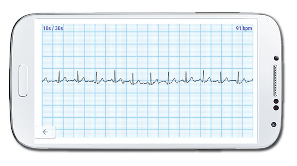
  • Digital ECG: Modern ECG machines use digital technology to produce high-quality recordings that can be stored electronically, making them easily accessible for review and analysis.
  • Telemedicine: ECGs can now be performed remotely, with the results transmitted to healthcare providers for real-time assessment, allowing for quicker diagnosis and intervention.
  • Machine Learning and AI: Artificial intelligence and machine learning algorithms are increasingly being used to assist in the interpretation of ECG data, improving accuracy and efficiency.
  • Wearable ECG Devices: Consumer-oriented wearable devices can record ECG data continuously, allowing individuals to monitor their heart health and share data with healthcare professionals.

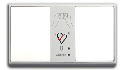
Ready to conduct your own EKG test at home?

The HeartCheck™ CardiBeat mobile ECG device can receive accurate electrocardiogram (ECG or EKG) readings in as few as 30 seconds that can then be sent instantly to a physician for medical review.